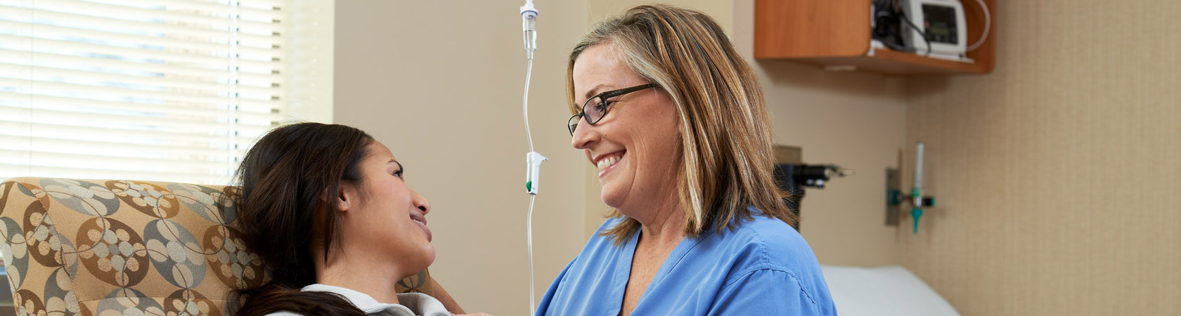
Botox & Dysport are treatments used to both prevent and treat wrinkles on the face that develop from repetitive muscle activity. They can be used in a natural way to enhance your appearance and help you look and feel like your best self.
How do Botox & Dysport work?
These are purified proteins that are used to relax the repetitive motion of overactive muscles in the face. The treatment is delivered through tiny injections directly into the facial muscles; Botox & Dysport then block the neurotransmitter that signals these muscles to contract. The result is a relaxed muscle that doesn’t make the same movement over and over again, which could otherwise lead to deep wrinkles. Botox & Dysport can soften existing wrinkles, but can also be used to prevent the development of lines on the face from repetitive motion.
Where do we use Botox & Dysport?
There are many areas of the face that can be treated with Botox & Dysport. The most commonly treated areas are the ‘elevens’ between the brows, the ‘crows feet’ that form outside your eyes, and the horizonal lines on your forehead.
How do I know which areas to have treated?
Prior to your treatment, we will have an honest and thorough discussion about your goals and the optimal aesthetic outcome. Only after this discussion will we determine the best unique treatment plan for you. Some people will treat just one area, and some will treat multiple areas of the face. Treatments are tailored to your skin and your goals, in order to give you a natural looking outcome.
What should I expect during my treatment?
After our initial consultation, it takes just a few minutes to perform the treatment. Minimal discomfort is managed with ice packs and most people have only minor redness following the treatment, which resolves in a few minutes. There is no significant downtime in most cases, and patients can return to work immediately following their treatment.
How long does Botox last?
Botox & Dysport may start to ‘kick in’ a few days after your treatment, but full results will be seen at 10-14 days after the visit. The results of your treatment will last on average 3-4 months.
Who performs the treatments at OMSC?
Lauren Sundick and Brooke Moss, also known as ‘The Skin Sisters,’ are experienced Physician Assistants specialized in Dermatology with over two decades of combined experience. They bring their love of aesthetics and patient relationships to our practice and are known for very naturalappearing outcomes.
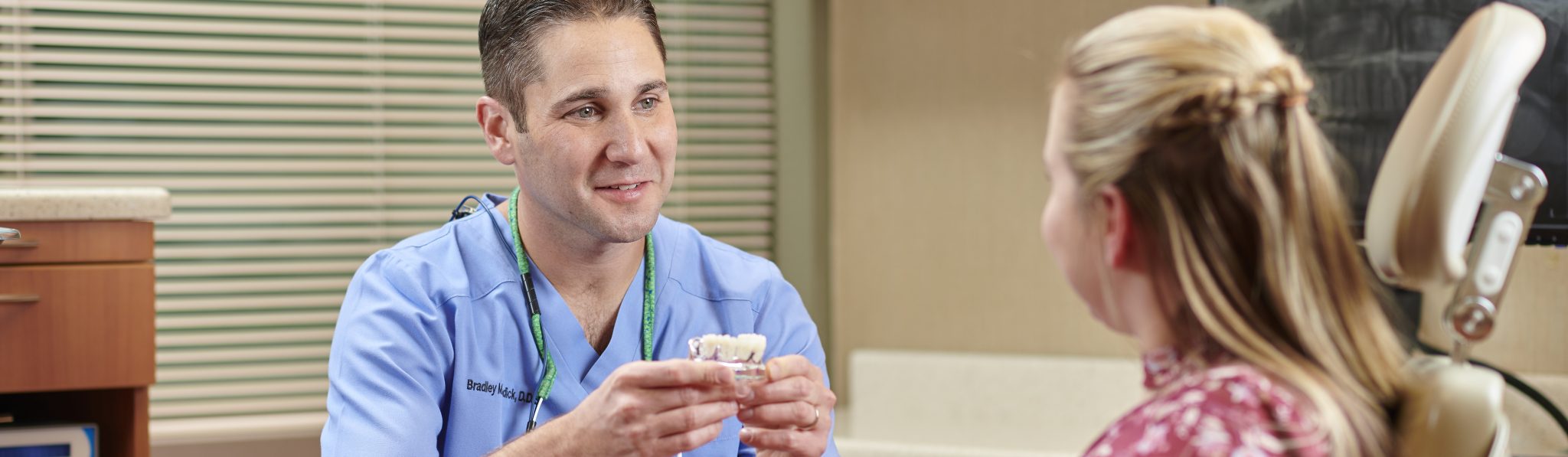
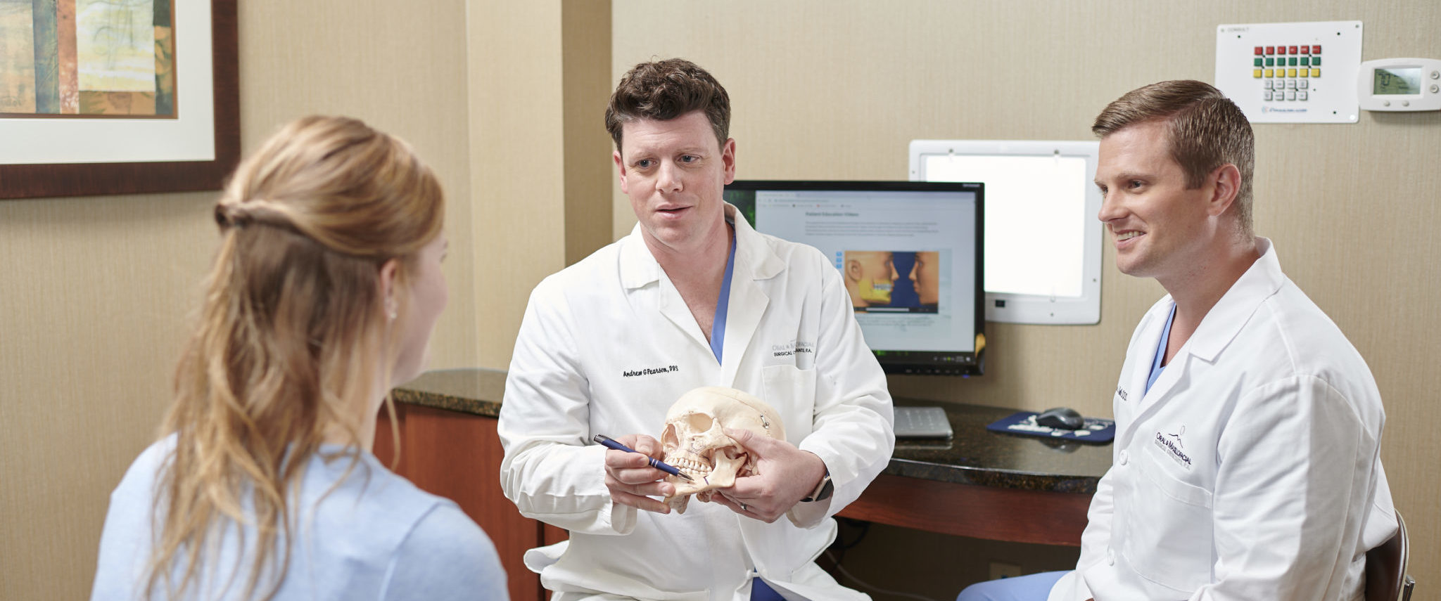
Similar to wisdom teeth, canine teeth sometimes have difficulty erupting and become impacted. Also called cuspids or eye teeth, they are usually the last adult teeth to erupt. Impaction of these teeth is often the result of dental crowding, lack of space, or failure of a primary/baby tooth to fall out. Canines are important teeth not only for chewing, but also contribute to speech, esthetics, and provide structural support for the surrounding hard and soft tissues.
Treatment for impacted canines is often easier and more successful the earlier it is identified. If these teeth do not erupt naturally, typically an orthodontist and oral and maxillofacial surgeon will collaborate to examine and determine the best treatment for each patient. This procedure typically involves the oral surgeon exposing the impacted tooth and placing a small orthodontic appliance on the tooth that will be used to guide the tooth into its proper position by the orthodontist. The position of the teeth and the degree of impact can vary greatly between individuals. The surgeons at Oral & Maxillofacial Surgical Consultants will help explain the findings and treatment options for each patient on an individual basis.
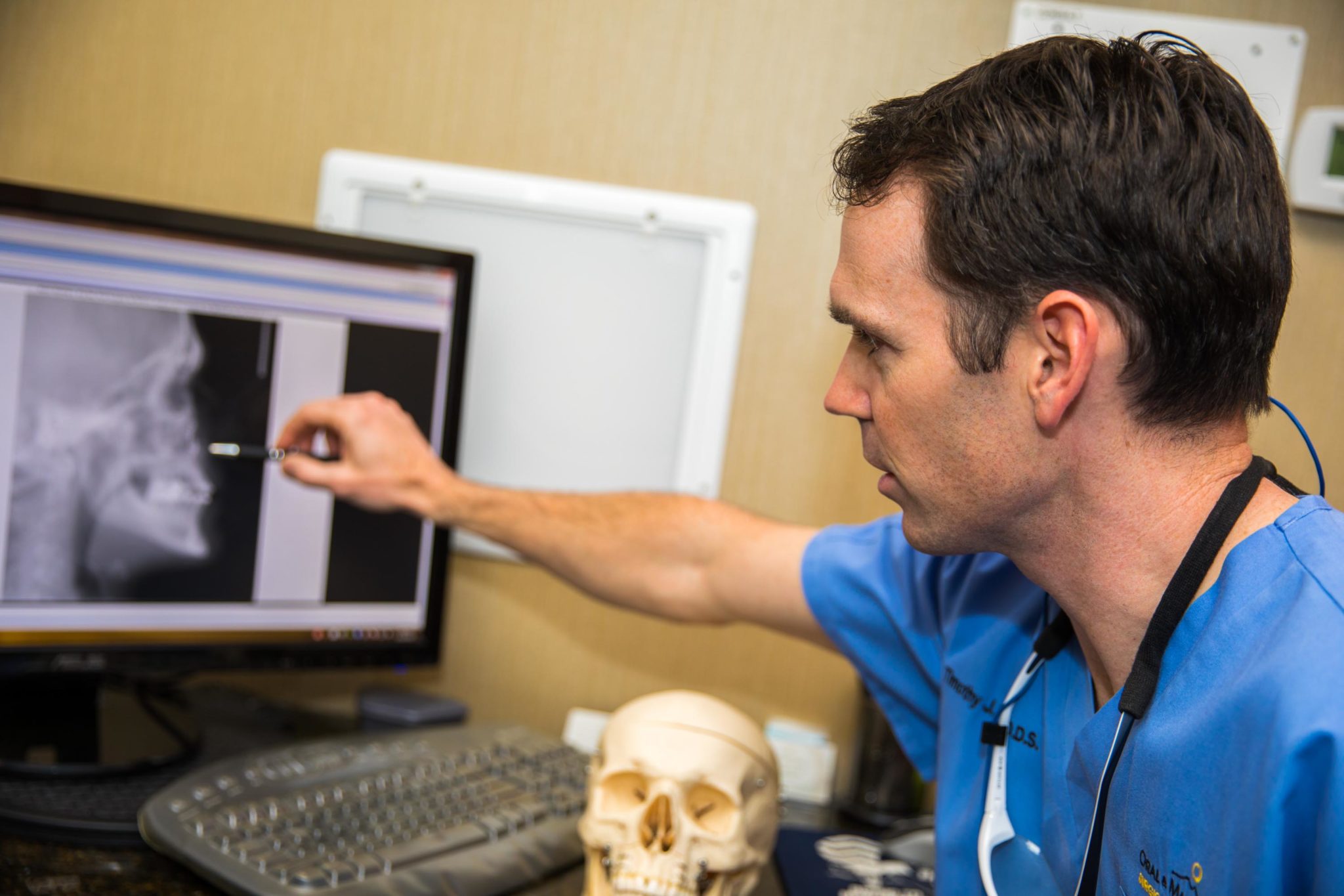
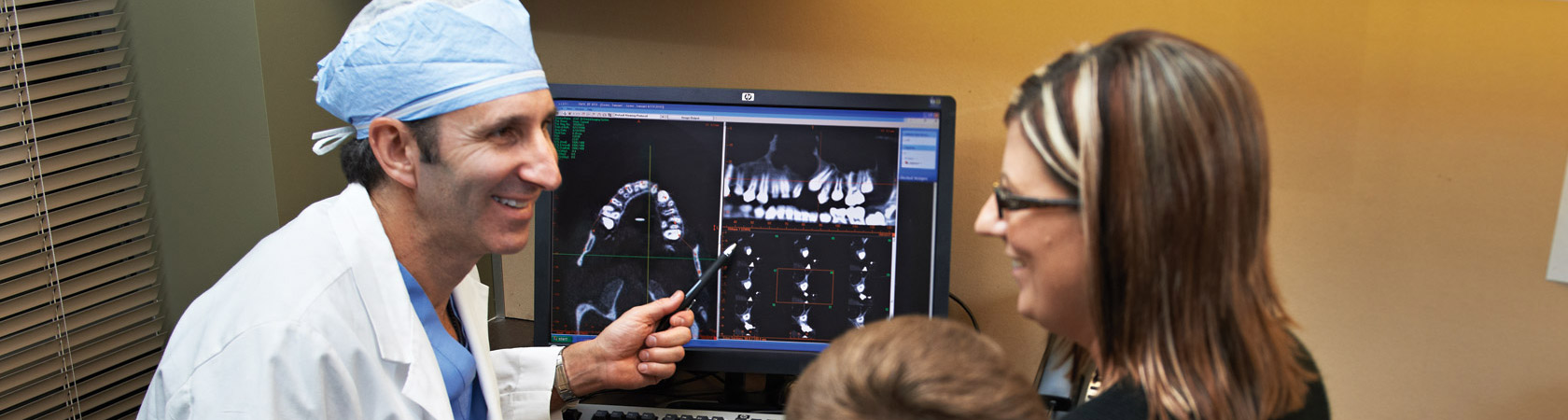
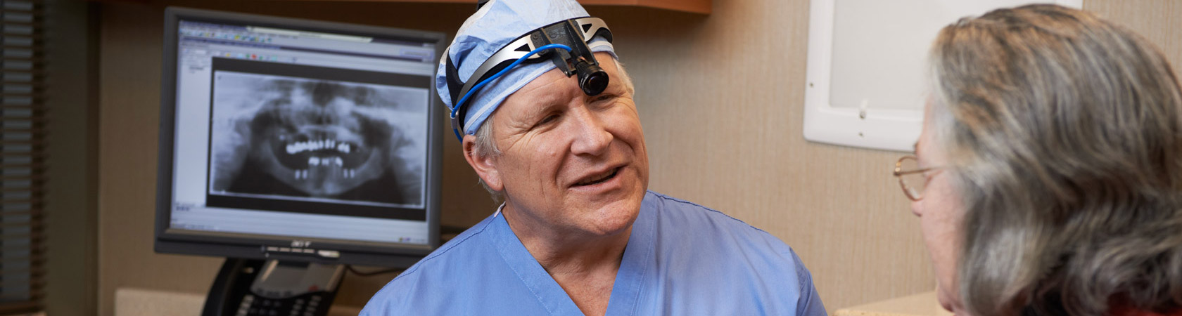
Oral and Maxillofacial Surgical Consultants has some of the most current and state-of-the-art cone beam 3-D dental imaging systems available. This type of technology provides high-definition, three-dimensional, digital imaging at a reduced cost and significantly less radiation than traditional medical CT scans. The images can be taken right in the office, removing the burdensome task of visiting a radiology center to have a CT scan obtained. This saves the patient time, money, and delays in treatment.
The technology’s 3-D, volumetric imaging system provides a complete view of all oral and maxillofacial structures, providing thorough diagnostic information possible. Our surgeons are able to more accurately diagnose and predictably treat patients than with conventional two-dimensional imaging. All aspects of the facial skeleton can be evaluated with high accuracy. Measurements are all one-to-one, with no magnification error.
Patients can view their CT scan right along-side their surgeon, allowing patients to better understand their anatomy. This helps patients better understand their diagnosis, treatment plan, and expected outcome.
OPTIMIZING PATIENT CARE:
Our surgeons believe that all patients deserve a high standard of care and this technology helps us meet that goal. Drs. Haas, Kurtzman, Pearson, Sundick, Neuner, McMahon, Spanel, Omlie, Ziegler, Afwerke and McGowan use cone beam 3D imaging to determine precise tooth position and the proximity of other vital structures, such as adjacent teeth, adjacent nerves, and other bony structures to ensure treatment is safe, accurate and effective.
Common Oral Surgery Procedures using 3D Imaging:
Impacted Teeth/Impacted Canine Teeth:
Three-dimensional cone beam imaging provides more surgical certainty when treating impacted teeth than when diagnosed with two-dimensional periapical or panoramic radiographs. The precise location of an impacted tooth and its exact relationship to the inferior alveolar nerve, maxillary sinus, or nasal cavity can be identified. This allows our surgeons to better explain the treatment and potential risks to each patient. Visualizing the anatomy more accurately reduces surgery and anesthesia time, which translates into a more safe, effective, and efficient surgery, leading to lessened discomfort and improved healing.
Implant Treatment planning and Implant Placement:
Three-dimensional cone beam imaging provides surgeons and patients with the best information available to develop a treatment plan for implant placement. It is an extremely diagnostic study tool for implant treatment planning in both the maxilla and mandible.
Vital structures and bone anatomy can be identified at the desired implant locations and measured with accurate, distortion-free images. Opposing teeth are visualized as well, which provides for better implant placement and overall prosthetic outcomes.
Orthognathic (jaw surgery) Treatment planning:
Three-dimensional cone beam imaging can provide a complete view of all oral and maxillofacial structures. This information can then be used to complete virtual surgical planning prior to orthognathic surgical procedure. By using virtual surgical planning, a surgeon can accurately visualize structures and assess proposed treatments prior to the actual surgery. This technology can help reduce surgery time and anesthesia time, which translates into a more safe, effective, and efficient surgery, leading to lessened discomfort and improved healing.
Other applications in dentistry:
Three-dimensional cone beam imaging technology has applications for every dental specialty, including:
The surgeons at Oral & Maxillofacial Surgical Consultants are experienced in the management of facial and dental trauma. We are trained and uniquely qualified to manage and repair injuries to the mouth, face, and jaws. Many dental and facial injuries result from sporting events, auto accidents, work-related accidents, home-related accidents, or acts of violence.
Our surgeons work with local hospitals and emergency rooms to treat facial injuries. Skilled treatment can lead to better outcomes in healing, eating, speaking, chewing, swallowing, breathing, and vision.
Drs. Haas, Kurtzman, Pearson, Sundick, Neuner, McMahon, Spanel, Omlie, Ziegler, Afwerke and McGowan recommend taking precautions to avoid any type of facial trauma, particularly in relation to sporting events and rigorous travel activities. It is always recommended for an individual to use a mouth guard and appropriate protection such as a helmet or facial shield when participating in any sporting activities that could cause injury to the face or teeth.
If a facial or dental injury does occur, seek medical attention immediately. Facial and dental injuries most often have the best prognosis when treated early and without delay. Request that the emergency room physician enlist the consultation of a local oral and maxillofacial surgeon. Oral & Maxillofacial Surgical Consultants will help manage acute emergency situations and help with additional treatments and patient rehabilitation when needed.
Soft Tissue Grafting
Soft tissue treatments are simple procedures designed to improve oral health, and often smile esthetics. Frenectomy and gingivectomy are two common soft tissue services routinely performed by our talented team at Oral & Maxillofacial Surgical Consultants. As highly skilled surgeons, our doctors utilize precision technique to deliver the best possible results for your family. We place comfort at the crest of each patient experience and offer minimally invasive laser treatments in many cases. Laser therapy with our LightScalpel® laser replaces scalpels and reduces the need for anesthetic and incisions. We also offer a range of anesthesia options, to ensure a pleasant positive treatment experience if anxiety or fear is a concern. No matter the needs or age of your family member, you can expect a caring chairside manner and exceptional quality care when undergoing frenectomy or gingivectomy treatment with one of our expert oral surgeons.
Gingival Grafting / Soft Tissue Graft:
Gingival grafts:
The gingival tissues are critical to a person’s overall oral health and help maintain healthy teeth, underlying bone, and create an aesthetic smile. Often a patient’s gingival tissues can be damaged from gingivitis, periodontal disease, trauma, and even too much tooth brushing. This gingival damage can result in recession (gum loss), leading to tooth sensitivity, bone loss, or even tooth decay. With gingival grafting, a soft tissue graft can be used to cover the exposed tooth roots or improve existing gingival tissues.
Soft tissue grafts and dental implants:
In addition to gingival grafts being used around existing teeth, these grafts can be used to optimize the success of a patient’s dental implant. Grafts may be put in place prior to an implant placement to improve the tissue or placed after to improve the esthetics of an existing implant. The timing of graft placement depends upon each patient’s unique needs.
There are four common types of soft tissue grafts:
Free gingival graft: A portion of tissue is removed from the roof the mouth and transferred to another area of the mouth.
Pedicle graft: A small flap of tissue is moved laterally to an affected area.
Subepithelial connective tissue graft: Commonly used to cover exposed roots. This is taken from small flap of tissue from the roof of the mouth.
Allograft matrix: This is donated human tissue that has been specially processed to be used as grafting material.
Your surgeon will discuss the best options for your unique circumstance.
TMJ Disorders
The temporomandibular joint (TMJ) allows a patient to open and close his or her mouth and aids with chewing, eating, speaking, and facial expression.
Temporomandibular joint disorders (TMD) can develop from trauma or injury, jaw clenching, parafunctional habits, or certain medical conditions. Disorders of the TMJ can result in pain or discomfort causing a decreased ability to open the mouth, pain when opening and closing the mouth, joint clicking/popping noises, earaches, and headaches. If a patient were to experience TMD symptoms, please contact Oral & Maxillofacial Surgical Consultants to set up an exam and consultation.
Drs. Haas, Kurtzman, Pearson, Sundick, Neuner, McMahon, Spanel, Omlie, Ziegler, Afwerke and McGowan are experienced in identifying and diagnosing a patient’s specific TMJ disorder. They will work with each patient to develop a treatment plan, which could include home care exercises, oral splints, physical therapy, anti-inflammatory or muscle relaxants, steroid injections, surgery, or a combination of treatments.
Through every step of treatment, the surgeons will work to ensure safe and effective care for each unique individual.
Platelet-Rich Plasma
Platelet-rich plasma (PRP) therapy is a safe, convenient procedure that can be completed in an office setting and used in conjunction with various oral and maxillofacial surgical procedures. Plasma is the liquid portion of the blood that transports red and white blood cells and platelets through the blood stream in a patient’s body. It is obtained from a patient’s own blood and placed into the patient’s oral surgical site to help increase the rate of healing.
Drs. Haas, Kurtzman, Pearson, Sundick, Neuner, McMahon, Spanel, Omlie, Ziegler, Afwerke and McGowan can use PRP in numerous clinical applications, including:
Bone grafting procedures prior to dental implants
Bone defect repairs
Fistula repair between the nasal sinus cavity and the mouth
Although most insurance companies do not cover PRP therapy, research studies indicate that about 70% of patients receiving PRP therapy show surgical site improvement. It is not always necessary, but your surgeon at Oral & Maxillofacial Surgery Consultants will provide you with a thorough list of safe and effective treatment options depending on your surgical needs to help provide safe and effective treatment.
The surgeons here at Oral & Maxillofacial Surgical Consultants
are dedicated to helping our patients fully understand the procedures
they need before they are performed. Please click through the lefthand
column below to find several informational videos about procedures
involving wisdom teeth, impacted canines, tooth and bone loss and
grafting, dental implants, and jaw surgery. As always, if you have any
questions or concerns, please contact our team.
A cleft lip and/or palate is a congenital (present from birth) facial deformity that can involve the lip, maxillary bone, and the nose. The treatment needs of this defect can vary from patient to patient and, therefore, are best managed by a team of specialists competent in this area. Oral and maxillofacial surgeons are ideally suited to perform various complex treatments regarding this condition. Our surgeons have been members of the Minneapolis Children’s Medical Center Cleft Lip & Palate Team for almost two decades and are dedicated to treatment of this condition. Our surgeons work closely with other specialists including orthodontists, otolaryngologists, speech therapists, prosthodontists, pediatric dentists, geneticists, pediatric surgeons, and primary care doctors. They provide treatment aimed at achieving the safest and most effective outcome.
The Surgery:
Our surgeons play an important role in the treatment of children and adults with cleft lip and palate. One of the common procedures performed by our oral surgeons is bone grafting to the maxilla when bone is not present due the cleft formation. This surgery takes place in a hospital operating room under general anesthesia. During this procedure, a bone graft will be placed within the alveolar cleft defect in the mouth and the surrounding gum tissue will be closed over the grafted area. Various bone grafting materials have been used for this procedure, but the “gold standard” is autogenous (from a person’s own body) bone that is obtained from the patient’s hip bone. The small bone graft removal from the hip bone and current placement of the bone graft within the cleft defect are completed during the same surgery. After the surgery is completed, the surgeons will guide each patient post operatively to ensure a safe and healthy recovery. The experienced surgeons at Oral & Maxillofacial Surgical Consultants will work closely with every patient and his or her care team to determine the best treatment options for each patient’s unique circumstance.
Anesthesia is defined as the loss of sensation with or without the loss of consciousness.
One of the specific skills involved in the training of an Oral and Maxillofacial surgeon is the administration of safe and effective anesthesia. During an oral surgeon’s training, he or she spends on average about 800 hours of training devoted entirely to administering various anesthesia to patients ranging from local anesthesia to general anesthesia. As Oral and Maxillofacial surgeons Drs. Haas, Kurtzman, Pearson, Sundick, Neuner, McMahon, Spanel, Omlie, Ziegler, Afwerke and McGowan are specifically trained in administering anesthesia that follows the most up to date safe and effective techniques and monitoring equipment, ensuring that each patient will be as comfortable and as pain free as possible.
Depending upon a patient’s medical history and the proposed surgical procedures various anesthesia techniques can be used. The most common techniques used by the Drs at Oral & Maxillofacial Surgical Consultants are:
Local anesthesia:
This type of anesthesia involves administering anesthetic such as lidocaine to numb the area where an oral surgical procedure is going to be performed. Local anesthesia is used is almost every single surgical procedure completed. This type of anesthesia is typically used for simple extractions, biopsies, and other minor procedures.
Nitrous Oxide: (laughing gas)
This type anesthesia involves the patient inhaling a combined mixture of oxygen and nitrous oxide through his or her nose to experience the effects. Nitrous oxide has long been used in the field of dentistry and oral surgery due to its ability to provide both a sedative and a analgesic effect. While a patient is receiving this medication, the patient remains conscious but remains in a more relaxed and calm stating during completion of a procedure. This medication is delivered concurrently with local anesthesia. Once the procedure is completed, the nitrous oxide is discontinued the effects of the gas are no longer present within 3-5 minutes. With this type of anesthetic, the patient is still able to eat and drinking normally prior to the procedure and have no driving restrictions post operatively.
Intravenous Sedation:
This type of anesthesia involves placing an IV into a patient’s vein typically on the back of the hand or in the middle portion of the arm where it bends. The patient additionally has several monitors attached to him or herself in order keep the patient safe during the procedure. During the administration of the type of anesthesia, three providers including the surgeon, a dental assistant and a nurse are present. Various different medications can be administered, and a detailed description of the medications will be discussed and reviewed with each patient at the time of consultation and procedure. Depending on the patient’s medical history and procedure, this type of anesthesia will be used concurrently with local anesthesia, and it can be used for almost any surgical procedure. It is commonly used during the removal of impacted teeth such as wisdom teeth or procedures that are more complicated such as bone grafting or dental implant placement.
General Anesthesia:
This type of anesthesia is administered to a patient in a hospital or outpatient surgical center. Depending on patient’s medical history or procedure needs, there are times when general anesthesia is necessary. This type of anesthesia is administered by an anesthesiologist and the patient is subsequently intubated with a breathing tube while the surgeon performs the procedure. Patient requiring more complication procedures such as orthognathic surgery, jaw reconstruction, or management of trauma or infections are examples of procedures completed with this type of anesthesia.
Drs. Haas, Kurtzman, Pearson, Sundick, Neuner, McMahon, Spanel, Omlie, Ziegler, Afwerke and McGowan primary goal when performing a procedure is the patient’s safety and well-being. After a thorough review of a patient’s medical history and treatment needs, the Drs at Oral & Maxillofacial Surgical Consults will ensure that each patient’s procedure is completed in the safest and most comfortable way.
When person’s jaws do not align properly or when teeth are unable to bite together properly, a patient may need corrective jaw surgery. This type of surgery involves surgically moving the upper jaw, lower jaw, or both jaws into a position that can improve a patient’s oral health, and even his or her systemic health. This procedure is completed in an operating room at hospital under general anesthesia. Oral and Maxillofacial surgeons are specifically trained in performing these types of procedures. This treatment is typically coordinated between an oral surgeon and an orthodontist. Jaw surgery can correct numerous minor and major skeletal and dental irregularities unable to be treatment with orthodontic therapy alone, allowing patients to experience improved chewing, speaking, and breathing. The surgeons at Oral and Maxillofacial Surgical Consultants, are well trained in performing various orthognathic procedures, and have extensive surgical experience to ensure an excellent and safe outcome.
Common Reasons for Jaw Surgery:
The following conditions indicate that you should seek consultation from an oral and maxillofacial surgeon to determine if orthognathic surgery would be recommended:
Speech or breathing problems
Chewing, biting, or swallowing problems
Chronic jaw pain
Protruding jaw
Open bite
Receding chin
Sleep apnea
Excessive wear on teeth
Your initial examination:
When a patient visits with one of our experienced surgeons for consultation, a review of his or her medical history, a thorough exam, and review of x-rays, will be completed. We are able to demonstrate to a patient with the use of three-dimensional models, proposed surgical treatments by using the most current computer imaging and software, for a better understanding the surgical process. Drs. Haas, Kurtzman, Pearson, Sundick, Neuner, McMahon, Spanel, Omlie, Ziegler, Afwerke and McGowan are dedicated to your safety and surgical success.
Developing your treatment plan:
Our surgeons will help to determine what kind of corrective surgical procedure will be needed and be most beneficial. A comprehensive care team that includes a restorative dentist, an orthodontist, and an oral and maxillofacial surgeon, will work to ensure the best treatment plan and outcome for each patient’s specific needs. The surgeons at Oral and Maxillofacial Surgical Consultants will ensure that every patient is well informed about the procedure, surgical outcomes and expectations, and the timeline of when surgery will be completed.
After jaw surgery: Healing.
Due the fact that each individual’s treatment plan and surgery is unique, each patient’s healing process will be different. Initially after surgery, a patient will typically stay in the hospital for 1-2 days after the procedure, in order for the surgeon to closely monitor and provide care as needed. Patients will then go home to continue his or her healing process. Typically, individuals do not return to work or school after completing this procedure for 1-2 weeks, but each case can vary due to patient age, procedure completed, school, sporting activities, extra circular activities, and occupation.
Diet: Patients will eat a soft, non-chew, predominately liquid diet for several weeks after the procedure. Common foods consumed after jaw surgery are: soup, smoothies, ice cream, apple sauce, yogurt, mashed potatoes, cottage cheese, and protein shakes. Foods to avoid would be those that are hard, crunchy, or sticky such as raw vegetables, raw apples, chips, crackers, peanuts, toast, bagels, and steak.
Once a patient is fully recovered and healed from jaw surgery, his or her teeth will fit together correctly. The jaw will be in a position that is more healthy, balanced and functional. Once fully healed, he or she can resume all normal daily activities and resume a normal diet without restrictions.
A detailed recovery plan outlining the recommendations, expectations, and restrictions, will be formulated and provided to each patient before and after the surgery has been completed, so as to ensure he or she feels confident about the entire process from start to finish. The surgeons at Oral and Maxillofacial Surgical Consultants, will make every effort to ensure that jaw surgery is as safe, comfortable, and effective as possible.
Oral health not only involves the teeth but also the tissues around them. The tongue, gums, and cheek tissues are areas that should be evaluated by your general DDS and potentially an oral and maxillofacial surgeon to check for any potential irregularities. The surgeons at Oral & Maxillofacial Surgical Consultants recommend everyone performs an oral cancer self-exam each month. A person’s risk of having oral cancer can be increased due to smoking, consuming alcohol, or using smokeless tobacco. Patients should have his or her dentist for perform an annual examination and may send their patient’s our clinic for treatment if needed.
To perform the monthly oral examination, each patient should examine his or her mouth with a bright light and a mirror, such as a bathroom mirror.
Examine the inside of the lips.
Examine the roof of the mouth.
Look inside the cheeks and around of the gums of the teeth.
Protrude the tongue and examine all of its surfaces including the top, sides, and underneath.
Feel for lumps in both sides of the neck, including under the jaw.
If a patient wears dentures, remove the dentures, and examine the tissues underneath.
Look for:
White or red patches/spots
Sores that easily bleeding, are painful or do not seem to heal
Abnormal lumps or thickened mouth tissues
Chronic sore throat or changes in voice
Difficult chewing or swallowing
Lumps on the neck
Early diagnosis and treatment of a potential pathologic process can make a significant difference in the treatment. If a patient has any of these symptoms or concerns, contact the doctors at Oral & Maxillofacial Surgical Consultants right away. Our doctors will perform a clinical exam and diagnose any problems. If a treatment is needed, Drs. Haas, Kurtzman, Pearson, Sundick, Neuner, McMahon, Spanel, Omlie, Ziegler and Afwerke are well-trained and will thoroughly explain the findings, and provide guidance through any treatment that may be needed.
Once a tooth has been lost due to dental decay or trauma, there can be changes to the surrounding bone that used to hold the tooth in place. If the tooth or teeth have been missing for an extended period of time, the underlying bone structure may be diminished. When too much bone has been lost, implants may not be able to be placed in the bone in its current state. Fortunately, with the implementation of various bone grafting techniques, implants can still be possible for most individuals. Bone grafting techniques allow the patient to regain bone that has been lost in order to have implants placed. Depending on the degree of bone loss, different bone grafting techniques may be necessary.
Bone Grafting What Is It And How Does It Work?
Typically, bone grafting can be categorized in the minor or major grafting.
-Minor grafting: The implant sites have areas of insufficient bone making ideal implant placement not possible. Small deficiencies can often be treated with a minimally invasive procedure that can usually be completed at the time of implant placement. This type of grafting will heal concurrently with your implant to ensure an optimal result.
-Major grafting: Large deficiencies in the jaw bone can be present due to long term tooth loss, trauma, infection, or congenital disorders. If the proposed implant site or sites do not have enough bone, various bone grafting procedures may be needed prior to implant placement. Bone grafting materials can vary from bank bone to bone from the patient’s own body, such as the posterior jawbone or the hip bone [see below for graft types]. Once the bone graft has been obtained, it is then placed in the proposed implant site, fixated in place, and allowed to heal. During this healing process, the bone graft material heals and integrates (fuses) to the surrounding bone underneath the soft tissue. This process makes the area suitable for implant placement. The bone graft healing can take up to six months or longer prior to implant placement.
Various materials can be used in bone grafting and can be categorized into four main areas:
-Autograft: Bone taken from one site in an individual’s body and moved to another site. This typically requires creating two surgical sites: one from the area where the bone graft is harvested and one where the bone graft is placed. For many oral surgery procedures, this bone is typically obtained from the back portion of the lower jaw from inside the mouth.
-Allograft: This is laboratory processed human bone (cadaver bone), from a deceased registered donor from a medical tissue bank. This type of graft acts as a framework or scaffolding for a patient’s body to grow and develop new bone over and within.
-Xenograft: This is laboratory processed bone that comes from an animal. It is typically bovine (cow) bone. This type of graft acts similar to an allograft and acts as a framework or scaffolding for a patient’s body to grow and develop new bone over and within.
-Alloplastic: These types of grafting materials are synthetic or laboratory made materials. These products are substituted instead of using real bone. One such substitute is Bone Morphogenic Protein, commonly referred to as BMP. These proteins occur naturally in the human body, and act to stimulate and regulate bone growth and healing. This material can be used to help regain bone that has been lost in an area where an implant is desired but is not amenable to implant placement in the current state.